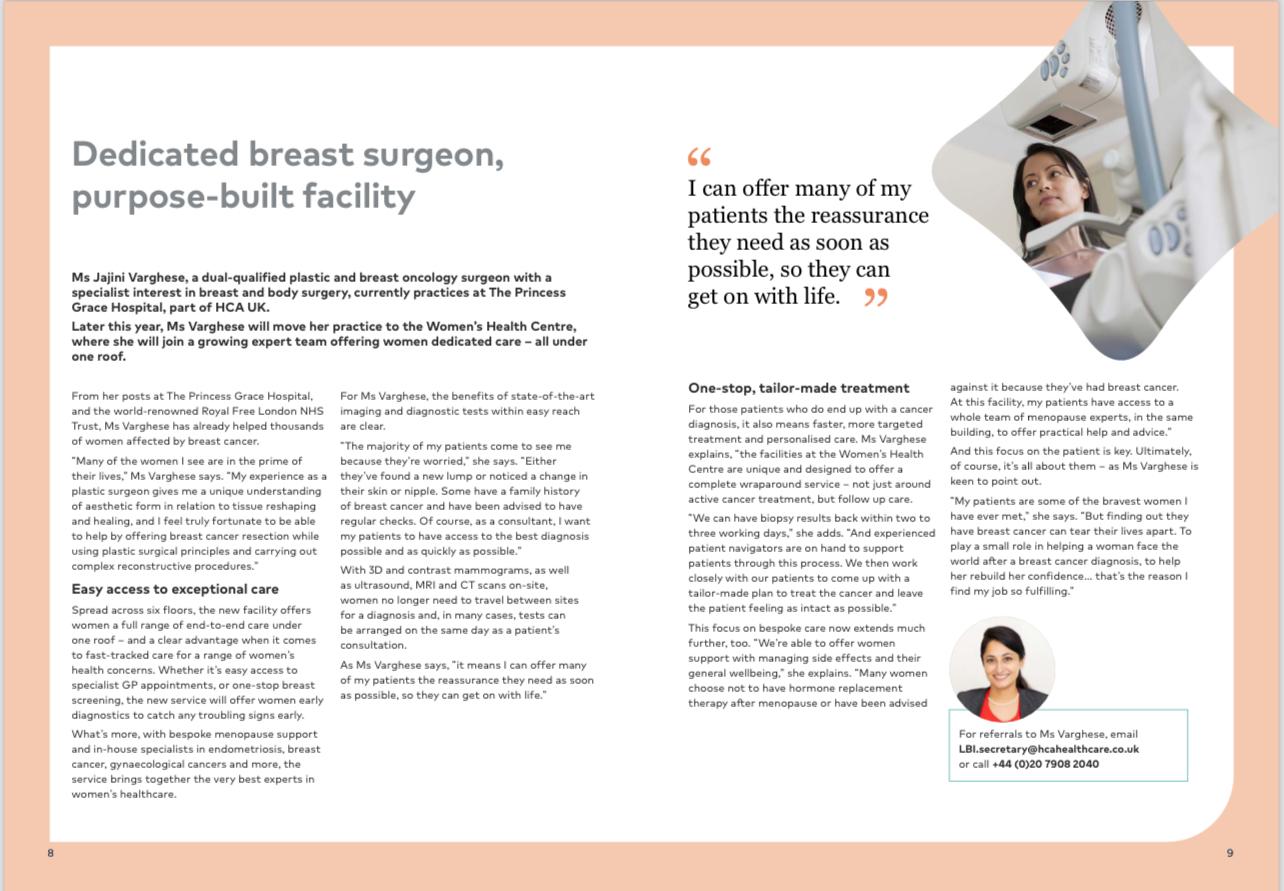
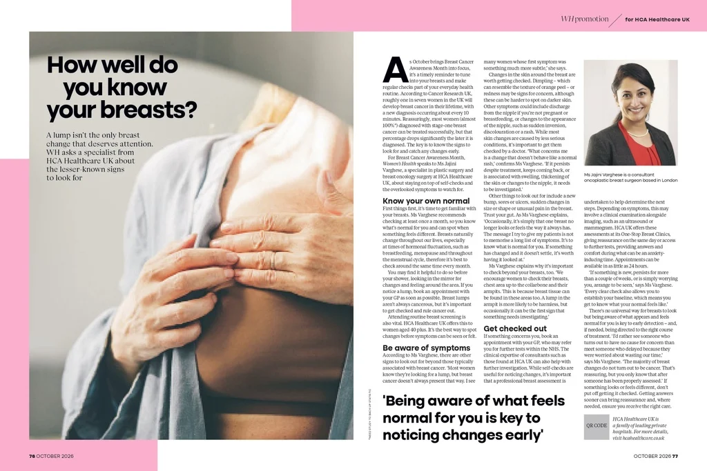
This is oncoplastic breast surgery explained the way I explain it across the desk in clinic. When a woman has just been told she has breast cancer, she usually asks two questions within minutes: will you be able to get it all out, and what will my breast look like afterwards? Oncoplastic surgery exists so that both questions are answered in one plan, with the first always taking priority over the second.
I have written this for women in the UK facing a diagnosis, or supporting someone who is. It covers what the surgery involves, how it compares with a standard lumpectomy and a mastectomy, who tends to be suitable, how margins work, and what recovery looks like for different techniques.
What Is Oncoplastic Breast Surgery?
Oncoplastic surgery combines two disciplines: oncology, removing the cancer safely, and plastic surgery, shaping the breast. The cancer operation comes first. Plastic surgery techniques are then used to keep, or rebuild, a natural breast shape.
The analogy I use most is a cushion. If you cut a piece out of one side and simply stitch the edges, you are left with a dent. In oncoplastic surgery, I remove the cancer with a margin of healthy tissue, then rearrange the remaining tissue so the breast still looks like a breast, perhaps a little smaller or slightly lifted.
It is not a single operation. The Association of Breast Surgery (ABS) and BAPRAS oncoplastic guidelines describe a family of approaches:
- Breast conserving surgery with local reshaping
- Therapeutic mammoplasty, using breast reduction or uplift patterns
- Chest wall perforator flaps, such as LICAP and TDAP
- Mastectomy with immediate or delayed reconstruction
- Symmetrising surgery on the other breast, where needed
Why Breast Conserving Surgery Shape Can Be a Problem
Breast conserving surgery, also called lumpectomy or wide local excision, removes the cancer and a rim of healthy tissue while keeping the rest of the breast. For many early cancers it is followed by radiotherapy, and the NICE guideline NG101 sets out how it should be offered alongside mastectomy, including the use of oncoplastic repair techniques where relevant.
The difficulty is proportion. Removing a 3 cm area from a large breast may leave little visible change. Removing the same amount from a smaller breast, or from the lower or inner part of the breast, can cause dimpling, a shift in nipple position or a clear difference between the sides. Radiotherapy can then make that distortion firmer and more noticeable over the following months.
Oncoplastic Surgery vs Lumpectomy: The Difference
The difference between oncoplastic surgery and a standard lumpectomy lies mainly in what happens after the cancer is removed. In a standard wide local excision, the cavity is closed simply and the tissue settles into the space. In an oncoplastic operation, tissue is deliberately moved or added to fill that space.
That can allow a larger volume to be removed while keeping a reasonable shape. A review in the Annals of Breast Surgery discusses how these approaches widen the group of women for whom breast conservation is realistic.
| Feature |
Standard lumpectomy |
Oncoplastic breast conserving surgery |
| Main aim |
Remove cancer with clear margins |
Remove cancer with clear margins, then reshape |
| Tissue removed |
Usually smaller volumes |
Can allow larger volumes |
| Closing the cavity |
Simple closure |
Tissue rearranged (displacement) or replaced (replacement) |
| Scars |
Often one shorter scar |
Often longer, sometimes reduction-pattern scars |
| Operating time |
Shorter |
Longer |
| Other breast |
Usually untouched |
Symmetrising surgery may be offered |
A standard lumpectomy remains a very good choice for many women, especially when the tumour is small relative to the breast. Oncoplastic surgery is not automatically better. It is the tool I reach for when a simple closure is likely to leave a poor shape.
Oncoplastic Surgery vs Mastectomy
A mastectomy removes the whole breast. It may be advised when the cancer is large compared with the breast, there are several separate areas of cancer, radiotherapy is not possible, or there is an inherited risk such as a BRCA variant. Some women choose mastectomy after full discussion, and that is a valid decision.
For some women who might once have been advised mastectomy only because a lumpectomy would have left too much deformity, oncoplastic conservation can be an alternative. Mastectomy also sits within oncoplastic practice, since reconstruction can be done at the same operation or later, using implants, your own tissue, or both. Our page on mastectomy with breast reconstruction mastectomy with breast reconstruction.
Am I Suitable for Oncoplastic Surgery?
Suitability is decided case by case, but some broad patterns help. You may be a candidate for volume displacement (such as therapeutic mammoplasty) if you have medium to larger breasts, some natural droop, and would accept a smaller breast. You may be a candidate for volume replacement (such as a LICAP or TDAP flap) if your breasts are small to medium, the cancer is in the outer or lower part, and you would like to keep your size.
Factors that may point elsewhere include:
- Cancer in several areas of the breast, or widespread DCIS
- A tumour that is very large relative to the breast, even after chemotherapy
- Reasons radiotherapy cannot be given
- Smoking, poorly controlled diabetes or a high BMI, which raise wound-healing risks
- A preference to avoid longer scars or surgery on the other breast
None of these is automatically a barrier. They are points to discuss openly.

Volume Displacement and Replacement: Oncoplastic Breast Surgery Explained Technique by Technique
Almost every oncoplastic breast conserving operation falls into one of two groups:
- Volume displacement moves the remaining breast tissue to fill the gap. The breast becomes smaller but keeps its shape.
- Volume replacement brings in tissue from nearby, usually the side of the chest, so the breast keeps more of its size.
Therapeutic Mammoplasty
Therapeutic mammoplasty for breast cancer treatment uses reduction or uplift patterns, planned so the cancer lies within the tissue being removed. The breast is reshaped and the nipple repositioned. Two scar patterns are common:
- Wise pattern: an anchor-shaped scar around the areola, down the front and along the fold. Suits larger breasts and bigger resections.
- Vertical scar: a lollipop-shaped scar around the areola and straight down. Avoids the horizontal scar and suits moderate reductions.
Because the treated breast becomes smaller, many women consider a matching reduction on the other side.
Chest Wall Perforator Flaps (LICAP and TDAP)
These use skin and fat from the side of the chest near the bra line, supplied by small vessels called perforators. LICAP (lateral intercostal artery perforator) flaps use vessels between the ribs. TDAP (thoracodorsal artery perforator) flaps use a vessel over the back muscle without taking the muscle itself. The donor scar usually sits along the bra line, and symmetrising surgery is often not needed.
Fat Grafting and Symmetrising Surgery
Fat grafting takes fat from the tummy or thighs by gentle liposuction and injects it into the breast. It is mainly used later to smooth dents, and often needs more than one session. Symmetrising breast surgery reshapes the other breast, either at the cancer operation or after radiotherapy changes have settled, which can make matching more predictable.
Wide Local Excision Margins and Re-excision
The first goal of any breast cancer operation is to remove all the cancer. Afterwards, a pathologist examines the edges of the removed tissue, the surgical margins, under the microscope. A clear margin means no cancer cells at the edge. NICE NG101 recommends offering further surgery if invasive cancer or DCIS is found at the margin.
Further surgery usually means a re-excision to remove more tissue from the relevant edge. Sometimes, particularly if margins are involved in several directions, mastectomy is the safer option. Your surgeon should tell you before your operation roughly how likely this is in your case, and what it would involve.
Oncoplastic reshaping makes re-excision more complex, because tissue has moved and the original cavity is no longer where it was. To manage this, we:
- Plan using your scans and biopsy, often with a marker or wire placed beforehand to locate the tumour
- Orientate the specimen and may send extra shave margins
- Place small titanium clips in the cavity walls before reshaping, so pathology, radiology and radiotherapy teams know exactly where the cancer was
Because larger volumes are often removed, oncoplastic operations may have similar or lower re-excision rates than simple lumpectomy in some series, though results vary, as discussed in the British Journal of Surgery (https://academic.oup.com/bjs/article/95/1/4/6156152). That is evidence about groups of patients, not a promise about any one person.
How the Decision Is Made: MDT and Radiotherapy Planning
In the UK, every breast cancer plan is reviewed by a multidisciplinary team (MDT) of surgeons, radiologists, pathologists, oncologists and specialist nurses, in the NHS and in reputable private practice. The MDT weighs the cancer’s size, type, grade and position; your breast size and shape; likely chemotherapy, hormone therapy or radiotherapy; and your own wishes.
Most women also have a sentinel lymph node biopsy under the arm at the same operation, to check whether cancer has spread to the nodes.
Post-surgery radiotherapy planning shapes the operation itself. Whole-breast radiotherapy is usually recommended after breast conservation, and wounds need to have healed first. The NICE breast cancer quality standard emphasises that women should be given information and choice about conservation, mastectomy and reconstruction. Variation in how services deliver this has been examined by the NHS Getting It Right First Time breast surgery programme.

An Illustrative Patient Journey
This is a composite created for illustration. It does not describe a real patient, and the details are deliberately typical rather than ideal.
“Anna” is 56. A screening mammogram finds a cancer in the lower outer part of her left breast, measured at 2 cm on ultrasound but closer to 3 cm on MRI, with some surrounding DCIS. That difference matters, and it takes a second MDT discussion to settle the plan.
She is offered two realistic options: a therapeutic mammoplasty with a vertical scar, or mastectomy with reconstruction. She leans towards mastectomy at first, worried about needing a second operation. After talking through her likelihood of re-excision and seeing where the scars would sit, she chooses the mammoplasty. She decides to delay surgery on her right breast until after radiotherapy, preferring one smaller operation now.
Her pathology shows one close margin. A re-excision is discussed, and she has it three weeks later as a day case. A small area of delayed healing at the scar junction pushes her radiotherapy back by about two weeks. A year on, she is considering a reduction on the other side and is still deciding. Her journey was neither straight nor tidy, which is common, and each step was her choice.
Oncoplastic Breast Surgery Recovery: What to Expect
Recovery depends on the operation. The ranges below are typical starting points for conversation, not targets, and your own team’s instructions come first.
| Procedure |
Typical hospital stay |
Desk work |
Driving |
Full activity and exercise |
| Volume displacement (therapeutic mammoplasty) |
Day case or 1 night |
Around 2 to 3 weeks |
Around 2 to 3 weeks |
Around 6 weeks |
| LICAP or TDAP flap |
Usually 1 night |
Around 2 to 4 weeks |
Around 2 to 4 weeks |
Around 6 weeks |
| Mastectomy with implant reconstruction |
1 to 2 nights |
Around 3 to 6 weeks |
Around 4 to 6 weeks |
Around 6 to 8 weeks |
| Mastectomy with own-tissue (e.g. abdominal) reconstruction |
3 to 5 nights |
Around 6 to 8 weeks |
Around 6 weeks |
Around 8 to 12 weeks |
Longer ranges apply if you have chemotherapy, a wound problem or a physical job. Drive only when you can wear a seatbelt comfortably and make an emergency stop. Most women wear a soft, non-wired support bra day and night for several weeks, and do gentle arm exercises, particularly after lymph node surgery.
Complications in Context
The most common issues after oncoplastic conservation are minor: seroma (fluid collection), bruising, and small areas of delayed healing, often where scars meet in a Wise pattern. Less common are infection needing antibiotics, bleeding needing a return to theatre, and fat necrosis, a firm lump of damaged fat that can feel worrying but is usually harmless and can be checked with imaging. Changes in nipple sensation are fairly common; loss of nipple blood supply is rare. Flaps carry a small risk of partial flap loss.
Smoking, diabetes and a higher BMI raise these risks. The main practical concern is that a wound problem can delay radiotherapy, which is why we look carefully at your health beforehand.
Expectations About Shape
Preserving shape does not guarantee symmetry. Breasts change with radiotherapy, weight and time. The ABS guidance recognises that oncoplastic treatment may include later adjustment operations, such as fat grafting or symmetrising surgery, and knowing this at the outset helps.
NHS and Private Routes
Oncoplastic surgery is available on the NHS, and many NHS breast units provide excellent care. Following a GP referral, you would be seen in a specialist breast clinic, and your treatment planned by the unit’s MDT. Some women choose private treatment for reasons such as choosing their surgeon, timing or continuity of care. The cancer principles, MDT review and margin standards are the same on either route.
For fees and how private treatment is arranged, please see our oncoplastic breast surgery page rather than relying on general figures. Breast Cancer Now offers free, independent support whichever route you take.
Useful questions for your consultation: Am I suitable for conservation? Which technique, and why? Where will my scars be? How likely is re-excision? How will this fit with radiotherapy? If you have a symptom but no diagnosis yet, a breast health assessment is the right first step.
Key Takeaways
- Oncoplastic surgery removes the cancer first, then rearranges or replaces tissue to keep a natural shape.
- Displacement suits larger breasts and makes them smaller; replacement suits smaller breasts and keeps their size.
- Clear margins come first, and re-excision is possible; ask how likely it is for you.
- Recovery and complications depend on the technique, and symmetry is never guaranteed.
Your Next Step
I hope having oncoplastic breast surgery explained in this way makes your options clearer. The aim is always the same: remove the cancer safely, then help the breast still look like a breast once treatment is over.
To discuss whether this approach suits you, visit our oncoplastic breast surgery service to arrange a consultation at Harley Street, the Royal Free or Hadley Wood Hospital, or contact our team with questions about referral.